
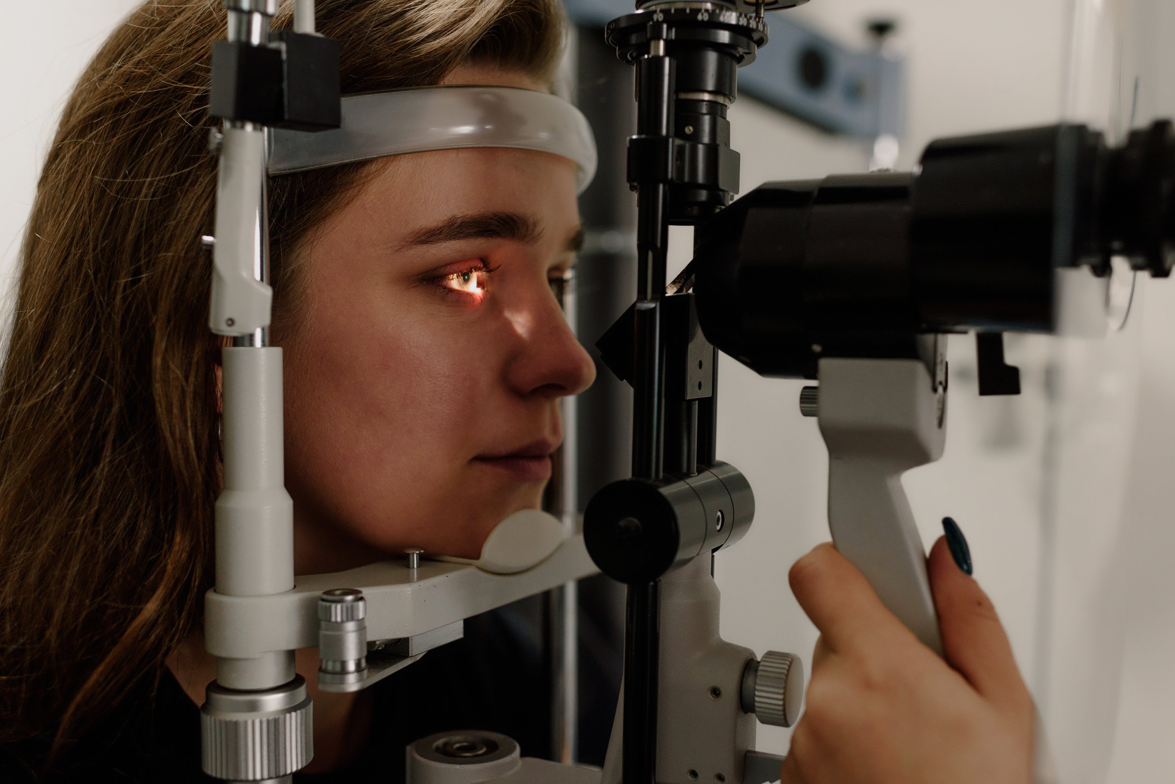
The invention is about real-time eye blink detection method, along with its placing at the core of human pupil
| Patent title | System analysis and automatic eye imaging selection deployable on ophthalmic devices |
|---|---|
| Thematic area | Health |
| Ownership | Università di Modena e Reggio Emilia, ALMA MATER STUDIORUM - UNIVERSITA' DI BOLOGNA |
| Inventors | Guido Borghi, GIOVANNI GIBERTONI, LUIGI ROVATI |
| Protection | National |
| Licensing status | Italy (IT) |
| Keywords | Pupillary Light Reflex; Ophthalmic Instrumentation; Eye Status Classification; Computer Vision-based Classification; Machine Learning; Deep Learning |
| Filed on | 15 December 2023 |
The system automatically selects images acquired from ophthalmic devices using AI-based visual techniques. It is primarily designed to enhance image capture activities in ophthalmics, providing support for ophthalmologists and/or eye surgeons when utilizing their tools, (when grounded in eye imaging).
The invention is a method for classifying eye features, accompanied by a device and a storage medium. This method is designed to address the challenge of poor classification of eye features in complex real-life context. The method involves an eye image acquisition and four types of labeled data by annotating eye image; this follows to the insertion of training data set in a neural network structure, designed to train an eye features classification model. Finally, the eye image to be classified is inserted into the eye feature classification model, and the output is the classification result.
The system is capable of real-time data processing directly on the embedded board, without the need to send data to a server or save images on a physical storage device, such as a hard disk or SSD. Furthermore, it doesn't require expensive hardware. The processing has been implemented to maximize computational efficiency, enabling real-time image classification with high accuracy. The performance peaks at 500 analyzed images per second for embedded systems and further reaches 2000 images per second on high-performance x86 CPUs. According to the state of the art, the system demonstrates a remarkable increase in processing efficiency, achieving high accuracy in classification (>95%)
Multiple low-cost applications can be observed in both the industrial and medical fields, primarily aimed at enhancing data acquisition activities or supporting the use of ophthalmic devices by physicians. The acquisition and processing techniques can also be applied to enhance existing ophthalmic systems, such as Optical Coherence Tomography (OCT), Visual Field Analyzer (VFA), Corneal Topographer, or pupillometry system.